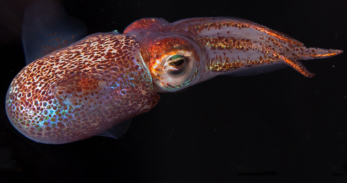
<![CDATA[New methods of fossil analysis have led a team of researchers from the University of Bristol and Virginia Tech to develop a method for detecting pigment in mammal fossils – something that could easily reveal the true colors of species that went extinct well in the past. Scientists were able to determine the shade of reddish brown of two extinct bat species dating to around 50 million years in the past. This represents the first time ever that fossil analysis has been able to determine the colors of an extinct mammal. The researchers said that the techniques they developed could be good for determining colors from animal fossils as ancient as 300 million years old, provided the fossils have been well-preserved. Virginia Tech geosciences doctoral student Caitlin Colleary, the study’s lead author, said in a statement published by the university that tissues from a wide variety of creatures, including ink from cephalopods like squids and octopuses, feathers from birds, hair and fur from mammals, and other tissue samples from aquatic and amphibious animals, all preserve melanin. This means that melanin has a high likelihood of being omnipresent in the fossil record, Colleary added, remarking that researchers can declare with confidence the color patterns of these long-extinct animals with a high degree of accuracy. The specific discovery made by the researchers is that cell organelles known as melanosomes – which are microscopic cellular structures that contain melanin – weren’t fossilized bacteria after all. Originally discovered and described by the University of Bristol’s Jakob Vinther, the senior author of the study just published this week, in 2008, fossil melanosomes have been used to investigate the evolutionary links between marine reptiles, and to identify the color animals like mammals and dinosaurs would have been in life. The research itself, which involved teams of scientists not just from the United States and the UK but also Denmark, Ethiopia, and Germany, was primarily undertaken at the University of Bristol, the location where Colleary originally worked with Vinther as a master’s student. Additional research was conducted at the University of Texas at Austin, which funded the project in part, along with the University of Bristol and National Geographic. Melanosomes are both chemically and physically distinct, as Vinther remarked in the press release. Melanosomes of different colors have different chemical makeups and physical shapes, something that was revealed during the research study thanks to the use of a time-of-flight secondary ion mass spectrometer. For more information: www.pnas.org Image courtesy of Wikimedia Commons user: Margaret McFall-Ngai ]]>
Color of Extinct Animals Revealed by Fossil Analysis